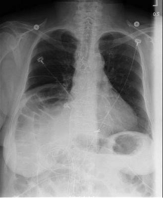
Chilaiditi's sign/syndrome

Ok this is the case---the elderly female diabetic was admitted for "pneumonia". She had right upper quadrant pain and on chest x-ray "an infiltrate". She was treated for pneumonia. After reviewing the chest x-ray and lab tests, the diagnosis was in question.
On further questioning of the patient, there was a 1 year history of constipation and generalized mild abdominal pain possibly worse the right upper quadrant. A diangosis of "gall bladder disease" was considered, but the ultrasound was normal as was the PIPDA. There was a history of "retained food" on a recent EGD. The diagnosis of "diabetic gastroparesis" was considered; but since she was a diabetic for less than 10 years and was only treated with an oral hypoglycemic, the diagnosis was in doubt.
Before dismissal she had a colonscopy and the prep made her "feel much better", but the scope could only get to the splenic flexure due to a "large redundant colon".
A chest x-ray was done prior to dismissal to follow up on the "pneumonia". The film is the one shown above. NOW the diagnosis of Chilaiditi's syndrome (or hepatodiaphragmatic colon interposition) is apparent.
NOW, after reviewing the symptoms of the syndrome, the correct diagnosis is more apparent.
While it is uncommon this film shows the BEST example of Chilaiditi's I have ever seen.
-Posted by Clay for Joe

3 Comments:
Ok this is the story---the elderly female diabetic is admitted for "pneumonia". She had right upper quadrnat pain and on cxr "an infilterate". She was treated for pneumonia. Reviewing the cxr and lab tests the dx was in question.
Further questioning of the pt. there was a history of 1 year of constipation and generalized mild abd. pain ? worse the ruq. A dx of "gall bladder dx" was considered, but the US was normal as was the PIPDA. There was a history of "retained food" on recent EGD. The dx of "diabetic gastroparesis" was considered. But being a diabetic for less than 10 years and only treatment an oral hypoglycemic, the dx was in doubt.
Before dismissal--she had a colonscopy and the prep made her "feel much better", but the scope could only get to the splenic flexure due to a "large redundant colon".
A cxr was done prior to dismissal to follow up on the "pneumonia". The film is the one shown. NOW the dx of Chilaiditi's syndrome(or hepatodiaphragmatic colon interposition} is apparent.
NOW reviewing the symptoms of the syndrome the correct dx is more apparent.
While it is uncommon the film shows the BEST example of the dx of Chilaiditi's I have ever seen.
I took the liberty of translating to English before posting so everyone could enjoy the case.
Thanks, blogmaster! How do you like the blister beetle?
2 more dxs today diskitis, and pityriasis rubra pilaris with pictures to follow.
Post a Comment
<< Home